Basic Concepts and Formulas in Microscopy
In order to realize the full potential of the optical microscope, one must have a firm grasp of the fundamental physical principles surrounding its operation. Important topics for understanding the microscope, such as resolution, numerical aperture, depth of field, image brightness, objective working distance, field of view, conjugate planes, and the useful magnification range, are discussed in the review articles linked below.
Review Articles
Innovations in Light Microscopy
A discussion of several new features available from the manufacturers.
Aberrations in Confocal Microscopy
How the trade-offs involved in objective design can affect confocal microscopy.
Basic Microscope Ergonomics
Modern microscopes feature user-friendly ergonomics that reduce fatigue and stress.
Conjugate Planes in Optical Microscopy
Image-forming and aperture planes form two independent focal sets in the optical train.
Depth of Field and Depth of Focus (日本語)
Fundamentals of the axial or longitudinal properties of microscope objectives.
Field of View
The eyepiece field diaphragm determines the diameter (size) of the microscope viewfield.
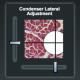
Coverslip Correction
Adjustments to the microscope objective to compensate for coverslip thickness variations.
Image Brightness
Brightness is proportional to either the square or the fourth power of the numerical aperture.
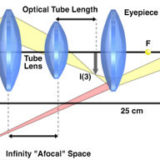
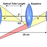
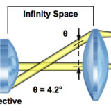
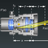
Infinity Optical Systems
These microscopes feature a parallel optical path between the objective and the tube lens.
Introduction to Microscope Objectives (日本語)
Objectives are responsible for image formation and the quality of images.
Laser Safety
Increased use of lasers demands attention to safety features in optical microscopy.
Microscope Objective Specifications (日本語)
The nomenclature and abbreviations inscribed on the objective protective barrel.
Modulation Transfer Function
A specification for the resolution performance of an optical microscope.
Multiple-Beam Interferometry
Microscopy using interference created between two independent light pathways.
Nikon CFI60 Optical System
The Nikon CFI60 specification for infinity corrected optics allows auxiliary components.
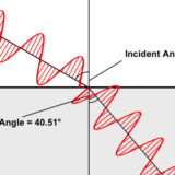
Numerical Aperture (日本語)
The ability of a microscope objective to gather light and resolve fine specimen detail.
Properties of Microscope Objectives
Discussion of numerical aperture, magnification, and aberration correction.
Refractive Index (Index of Refraction) (日本語)
The ratio of the speed of light in a vacuum to that in the imaging medium of a microscope.
Resolution (日本語)
The resolving power of a microscope is the most important feature of the optical system.
Specialized Microscope Objectives
Phase contrast, DIC, fluorescence, darkfield, Hoffman, and reflected light.
The Microscope Optical Train
The optical train consists of an illuminator, condenser, specimen, objective, and detector.
Two-Beam Interferometry
Microscopy using interference created between two independent light pathways.
Useful Magnification Range
Selecting the best combination of objective and eyepiece for the numerical aperture.
Water Immersion Objectives
Highly corrected objectives introduced for applications involving living cells and tissues.
Working Distance and Parfocal Length (日本語)
The distance between the objective front lens or the nosepiece and the specimen.
Linear Measurements (Micrometry)
A comprehensive review of eyepiece reticles and stage micrometers for measurements.
Interactive Tutorials
-
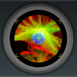
Astigmatism
An off-axis aberration manifested as a point appearing to be a line or ellipse.
-
Chromatic Aberration
Explore axial and lateral chromatic aberrations seen in an optical microscope with this interactive tutorial.
-
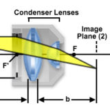
Condenser Image Planes
Examine the relationship between field and condenser diaphragm image planes.
-
Conjugate Planes
Use this tutorial to explore the image-forming conjugate planes in an optical microscope.
-
Coverslip Correction Collars
Investigate how internal lens elements in a high numerical aperture dry objective may be adjusted to correct for fluctuations in coverslip thickness.
-
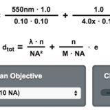
Depth of Field Calculator
Calculate the depth of field for popular objectives with this interactive tutorial.
-
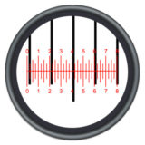
Eyepiece Reticle Calibration
Explores calibration of various eyepiece reticles using a stage micrometer and demonstrates how the reticle can then be employed to determine linear specimen dimensions.
-
Field Curvature
Common aberration caused by the spherical surface of lens elements.
-
Field of View Diameter
Exploring the effect of varying the field of view size on the viewable specimen area.
-

Focus and Alignment of Mercury and Xenon Arc Lamps
Explore alignment and focusing of the arc lamp in a mercury or xenon burner, which simulates how the lamp is adjusted in a real microscope.
-

Geometrical Construction of Ray Diagrams
A popular method of representing a train of propagating light waves.
-
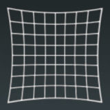
Geometrical Distortion
Termed pincushion or barrel, geometrical distortion often occurs in stereomicroscopy.
-
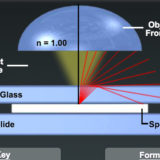
Immersion Oil and Refractive Index
The refractive index is critical in determining the working numerical aperture of an objective.
-
Infinity-Corrected Microscope Conjugate Field Planes
Objective light is focused to infinity and the tube lens forms the image at its focal plane.
-
Microscope Alignment for Köhler Illumination
Learn how to adjust a microscope to examine specimens in Köhler illumination.
-
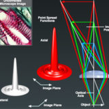
Microscope Conjugate Field Planes
The geometrical relationship between image planes in the optical microscope.
-
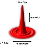
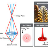
Numerical Aperture and Image Resolution
Explore how objective numerical aperture size influences Airy disk properties.
-
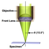
Numerical Aperture Light Cones
Numerical aperture is a measure of the highly diffracted light rays captured by the objective.
-
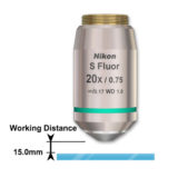
Objective Working Distance
Examine the range of working distance values available in modern Nikon CFI60 objectives.
-
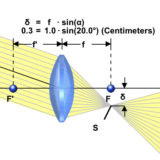
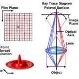
Perfect Lens Characteristics
An ideally corrected lens that is free of aberration and focuses light to a point.
-
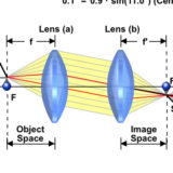
Perfect Two-Lens System Characteristics
Explore how off-axis oblique light waves pass through a two-lens system.
-
Photomask Reticle Operation
Practice adjustment of the photomask reticle in a focusing eyepiece.
-

Proper Microscope Posture
Explore proper head and back posture for microscope observations.
-
Refraction of Light
Examine how refractive index varies with the dispersion of properties of different materials.
-
Stereomicroscope Optical Pathways
Examine the light pathways in a Nikon stereomicroscope.
-
Specimen Contrast Enhancement with Apodized Phase Plates
Examine a variety of specimens using traditional and apodized phase contrast microscopy.
-
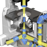
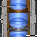
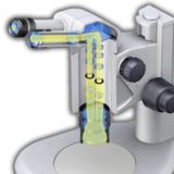
Inverted Microscope Optical Pathways
Examine an animated cut-away diagram of a modern inverted microscope.
-
Tube Lens Focal Length
Infinity optical systems feature tube lengths between 200 and 250 millimeters.
-
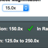
Useful Magnification Range
Determine if objective-eyepiece combinations are in the useful magnification range.
-
Viewing and Projection Eyepieces
A discussion of projection and viewing eyepiece systems.
Selected Literature References
Autofluorescence
Natural fluorescence in plants and animals that can also be introduced by fixatives.
Confocal Microscopy
Imaging with laser excitation and pinhole detection of fluorescence.
Differential Interference Contrast (DIC)
Contrast enhancement using interference of polarized light wavefronts.
Fluorescence Microscopy
Examining specimens labeled with molecules that absorb light and emit fluorescence.
Microscope Optical Systems
Information on the basic properties of microscope and camera optics.
Phase Contrast Microscopy
Using refractive index variations to produce optical contrast in transparent specimens.
Polarized Light Microscopy
Observing birefringent specimens using cross-polarized illumination.
Specimen Contrast in Microscopy
Examine the origins of contrast in a wide spectrum of specimens.
Spinning Disk Microscopy
Excellent technique for high-speed imaging of living cells in real time with a CCD camera.
Stereomicroscopy
Long working distance, low magnification microscopes for stereoscopic observation.
Contributing Authors
Kenneth R. Spring - Scientific Consultant, Lusby, Maryland, 20657.
Hiroshi Komatsu - Institute for Materials Research, Tohoku University, Sendai, Japan.
Martin L. Scott - Scientific Imaging, 231 Vassar St., Rochester, New York, 14607.
Stanley A. Schwartz - Nikon Instruments Inc., 1300 Walt Whitman Rd., Melville, New York, 11747.
Thomas J. Fellers, Kathleen E. Carr, Matthew Parry-Hill, and Michael W. Davidson - National High Magnetic Field Laboratory, 1800 East Paul Dirac Dr., The Florida State University, Tallahassee, Florida, 32310.
Share this page:


